

MRI, CT, and PET data visualized using 3D Slicer using direct volume rendering and indirect volume rendering.
All data used is sample.
Created as part of Master's course in Volumetric Visualisation at the School of Simulation and Visualisation, Glasgow School of Art.
Pelvic fracture: PRE + POST surgery
Comparison of pelvic fracture pre-surgery with the results of the surgery.
Indirect volume rendering (iDVR) of pelvic fracture pre-surgery. Post-surgery pelvic rendered directly (DVR) with decreased opacity to show pins, segmented as iDVR.
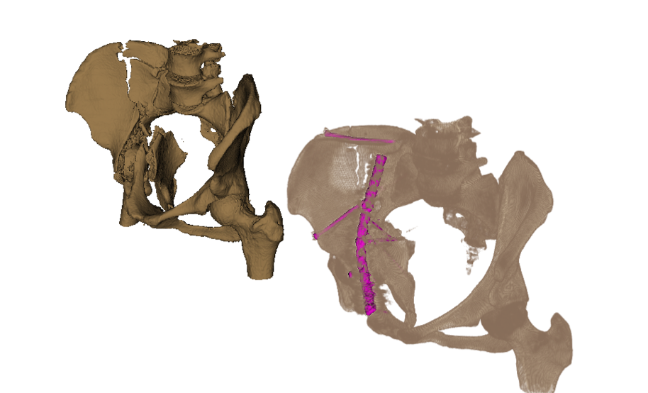
Comparison of pre- and post-surgery pelvis
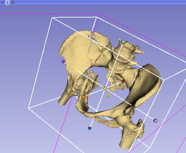
iDVR of pelvic fracture
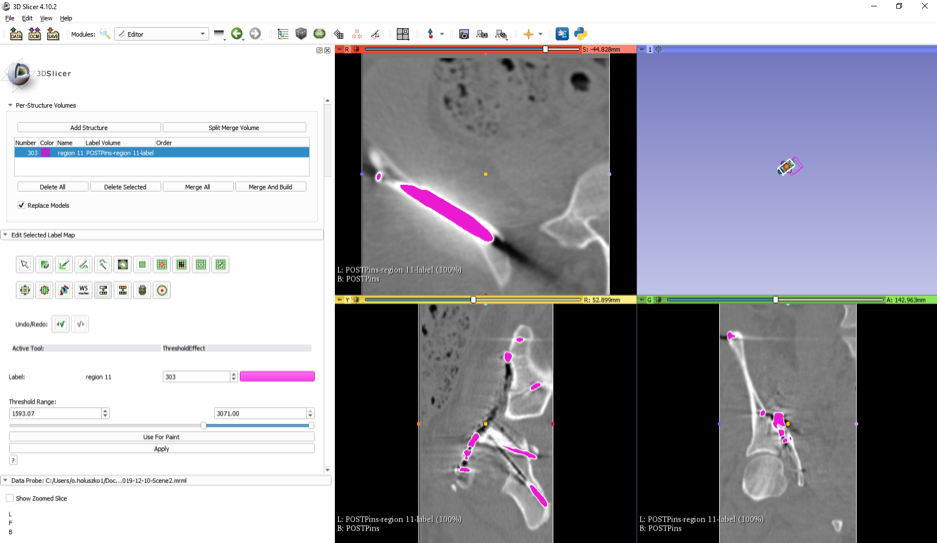
iDVR of pins in pelvis after surgery
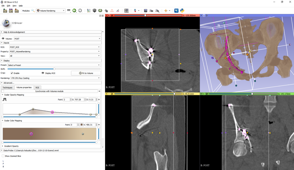
DVR of pelvis post-surgery to show pins
Brain tumour: multiple datasets
Localizing the brain tumour and showing it in its surrounding areas.
Aligned three datasets (MRI, CT, PET). Used iDVR of MRI to render the tumour, ventricles, and brain. Used DVR of CT to show the braincase.
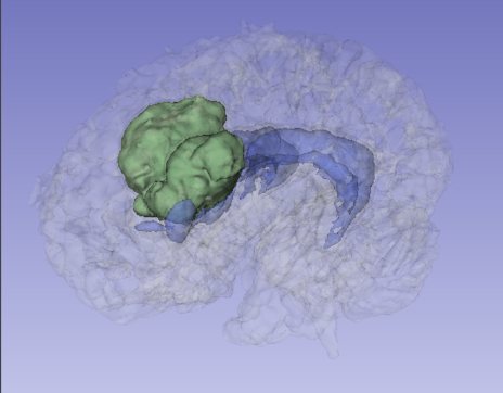
iDVR of tumour, ventricles, and brain, with adjusted opacities
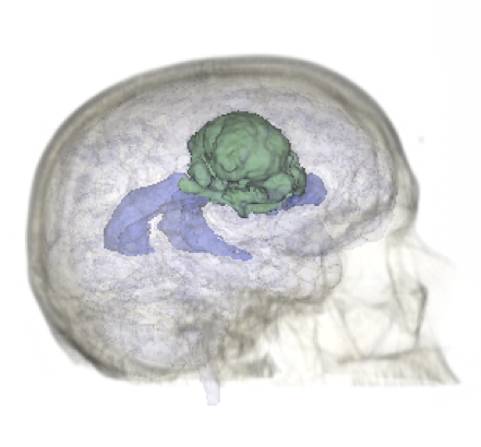
DVR of PET to localize brain tumour within skull
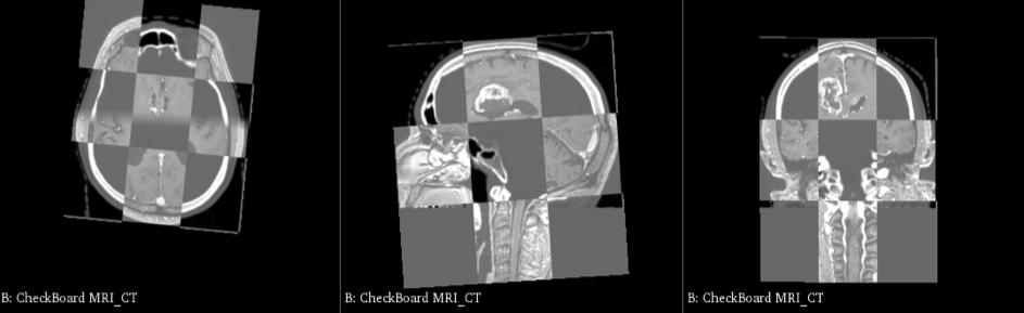
Aligning MRI and CT datasets
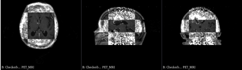
Aligning MRI and PET datasets
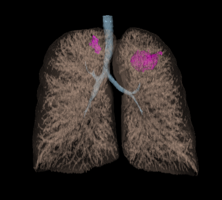
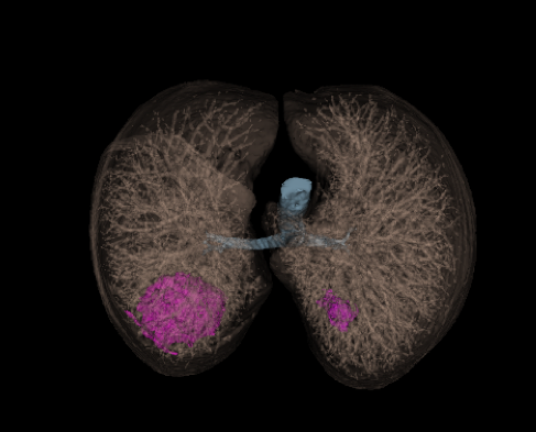
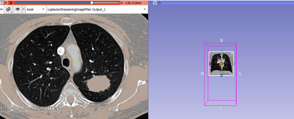
Lung tumours
Showing the tumours within the lungs and determining their respective volumes.
iDVR of the lungs, trachea, and the tumours to localize them in the body. Carefully segmented since the isodensities of the tumours were closely related to surrounding structures.
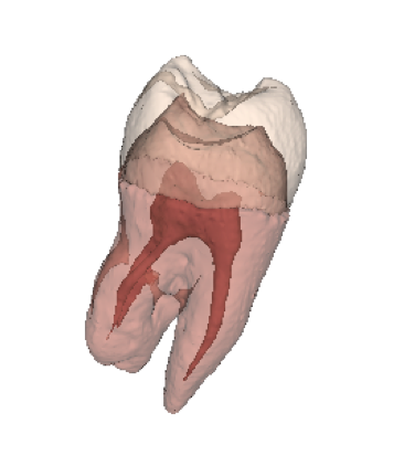
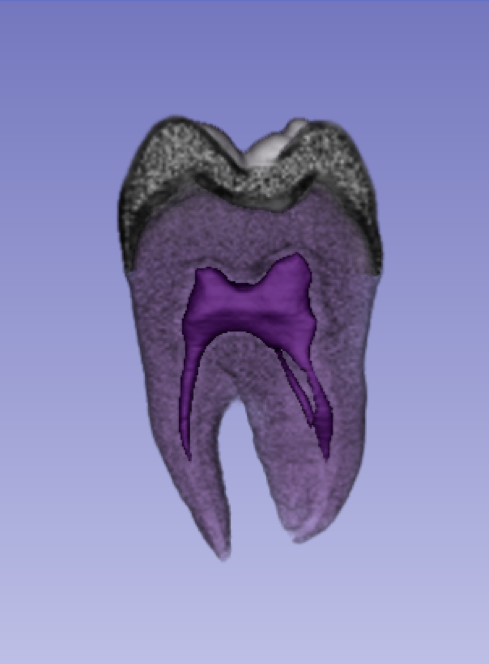
ANATOMY OF A TOOTH
Showing the internal structure of a tooth for educational purposes.
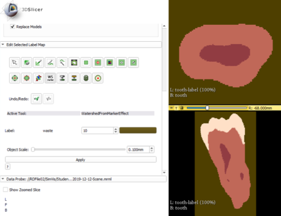
Used both iDVR and DVR to generate models of the tooth to show its internal structure and overlapping layers.
View more 3D Work
Anatomy of FGM/C Webapp / Anatomical 3D Models / Volumetric Visualisation / Krebs Cycle Game